Abstract
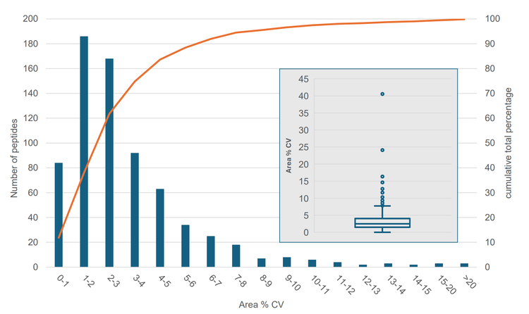
This technical note describes the fast and sensitive quantitation of a large panel of peptides in human plasma using the SCIEX 7500+ triple quadrupole mass spectrometry system. Peptide quantitation is well-established on triple quadruple platforms and is used extensively for biomarker research1. To ensure accurate quantitative performance, typically a minimum of two Multiple Reaction Monitoring (MRM) transitions are used for each peptide, which limits the number of peptides in the assay. The speed of the SCIEX 7500+ system maximizes the number of concurrent MRMs, which enables the analysis of very large panels of peptides. A multiplexed assay was developed with 2,836 multiple reaction monitoring (MRM) transitions for 709 peptides, both native and heavy-labeled, from 530 human plasma proteins using a 20-minute microflow liquid chromatography (LC) gradient. Using fast MRM scanning (combined dwell and pause times of 3.5 msec per MRM transition) on the SCIEX 7500+ system, >97% of the monitored labeled peptides were quantified with area coefficient of variation (CV) <10%, and the median CV across all transitions was 2.5%.
These results demonstrate the industry-leading performance of the SCIEX 7500+ system for accurate and precise absolute quantitation of large peptide panels, making it the ideal system for disease biomarker research.
Key features of large panel peptide quantitation using microflow LC and the SCIEX 7500+ system
- Faster analysis: Ultra-fast MRM acquisitions enabled by the SCIEX 7500+ system, with combined dwell and pause times as low as 3.5 msec per MRM transition, allows for fast multiplexed quantitative assays of large analyte panels
- Improved quantitative performance: Next-level quantitative precision and accuracy demonstrated using >2,800 MRM transitions to quantify 709 tryptic peptides in human plasma using a 20-minute microflow LC gradient, where >97% of the heavy labeled peptides had area CVs <10% and the median CV across all peptides was 2.5%
- High sensitivity: 5x less sample loadings required for the SCIEX 7500+ with microflow LC using a 0.15 mm ID column at a flow rate of 1.5 µL/min when compared to analytical flow LC
Introduction
After the discovery of potential protein biomarkers using unbiased proteomics analysis, verification of these candidate biomarkers in large numbers of samples is required. These targeted assays are typically performed using multiple reaction monitoring (MRM) with a triple quadrupole mass spectrometer, which provides high sensitivity and specificity for detecting low abundance proteins through proteotypic tryptic peptides1. Heavy-labeled proteotypic peptides are often spiked in for absolute quantitation, whereby protein concentrations are measured by comparing the response of the labeled peptide with the response of its native, light, counterpart. Throughput is essential as large numbers of samples need to be analyzed before a clinical application is possible. In this technical note, we demonstrate how the SCIEX 7500+ system provides excellent quantitative precision using only a 3.5 msec combined pause and dwell time. Scheduling MRM transitions with a 60-second window around the expected retention times allowed for the quantitation of 709 light/heavy peptide pairs with a 20-minute microflow LC gradient run. Using a 0.15 mm ID microflow column at 1.5 μL/min flow rate increases sensitivity while still offering a robust method.
Methods
Samples and reagents: Human pooled plasma K2EDTA was acquired from BioIVT. Top 14 Abundant Protein Depletion Midi spin columns from Thermo Fisher were used for plasma depletion. Trypsin/Lys-C protease mix was purchased from Promega. PQ500 heavy-labeled peptides were purchased from Biognosys.
Sample preparation: Depletion of the top 14 most abundant proteins was performed with the depletion spin columns using the manufacturer's protocol. The remaining proteins were digested following a filter-aided sample preparation (FASP) protocol described in the literature.2 After drying the tryptic peptides, they were dissolved with 0.1% formic acid in water to a concentration of 0.88 μg/μL, and PQ500 peptides were spiked in. The standard spike-in ratio of the manufacturer is 4 μL PQ500 mix to 12 μL of the (depleted) plasma digest diluted to an estimated protein concentration of 0.88 μg/μL, which was equivalent to 1.06 μL plasma. This ratio is referred to in this technical note as "1x". Additional samples were prepared with the same plasma digest concentration but PQ500 peptides at 0.05x, 0.25x, and 2x ratios to determine linearity around the 1x ratio spike in amount. The higher sensitivity from using microflow LC allowed for a further 4x dilution with 0.1% formic acid before injection. In addition, the amount injected per analysis was also lower than recommended (2 μL instead of 3 μL). For this technical note, only non-alkylated peptides, i.e., peptides without cysteines, were analyzed.
Chromatography: The samples were analyzed using a Waters ACQUITY M-Class system in trap and elute nanoflow LC mode. A Waters nanoEase M/Z Symmetry C18 100 Å, 5 μm, 180 μm x 20 mm trap column was used in combination with a Phenomenex Kinetex XB-C18 100 Å, 2.6 μm, 0.15 mm x 15 cm microflow LC column. 2 μL sample was loaded on the trap from a 10 μL loop using 1 minute of loading at 10 μL/min of 0.1% formic acid in water. A 20-minute gradient at 1.5 μL/min from 1-28% mobile phase B was run for the separation, using 0.1% formic acid in water as mobile phase A and 0.1% formic acid in acetonitrile as mobile phase B. The column and trap were washed at 80% mobile phase B for 1.5 minutes and re-equilibrated at 1% mobile phase B for 6 minutes. The column temperature was maintained at 40°C. All analyses were performed in triplicate.
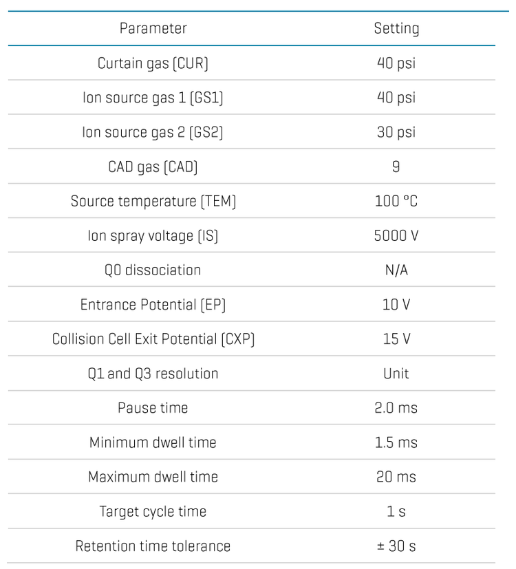
Mass Spectrometry: All analyses were performed using the SCIEX 7500+ system with an OptiFlow Pro ion source with micro E Lens™ and microflow probe with low micro electrode. Two fragments for each peptide were selected for optimal quantitation with Skyline software, using Zeno SWATH DIA data previously acquired on the ZenoTOF 7600+ system for the PQ500 peptides in a plasma digest3. Collision energies were calculated using the dynamic collision energy equation for peptides integrated in the SCIEX OS software. Ion source and MS method parameters for the final scheduled MRM method used are listed in Table 1.
Data processing: Data were processed using the Analytics module of SCIEX OS version 3.4. The two selected fragment ion signals for each heavy or light peptide were summed for quantitation and integrated using the Autopeak integration method.
Scheduled MRM method optimization
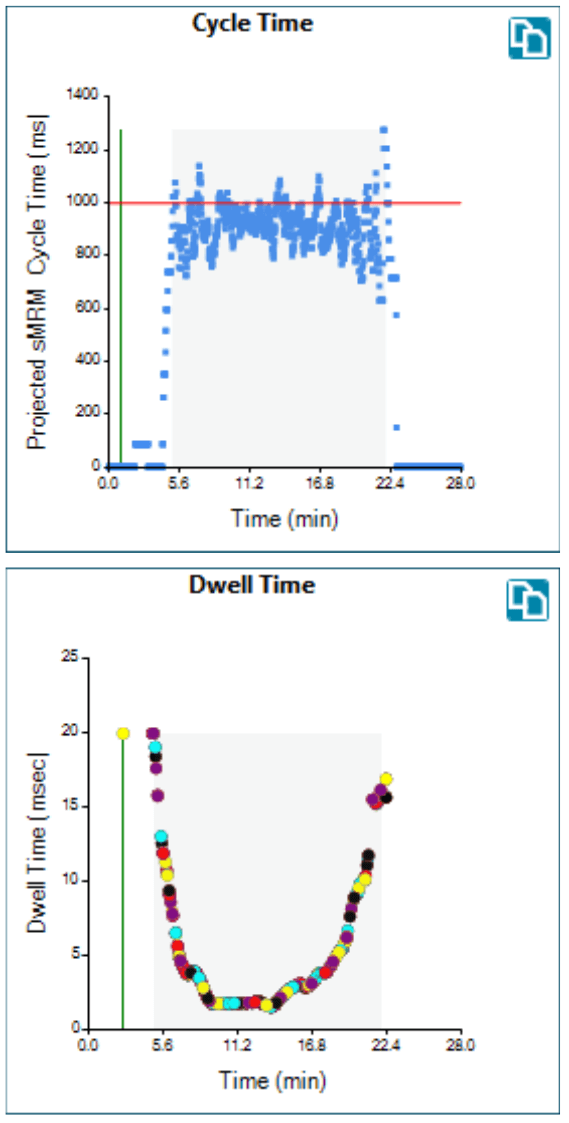
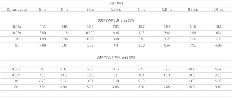
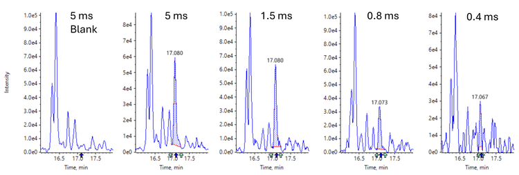
An experiment was performed with two lower abundant heavy peptides to optimize the dwell time, using an MS method with the same MRM transitions at different dwell times. Table 2 lists the peak area CVs at different dwell times and spike-in amounts, and Figure 3 shows the XICs for the sum of the transitions of one of the two peptides at different dwell times. Based on the observed CVs and XICs, a minimum dwell time of 1.5 ms, for a combined pause + dwell time of 3.5 ms, was selected for the scheduled MRM method. The optimal pause time for peptides, representing the time between two MRM experiments, was 2 ms. At lower pause times, the MRM signal can be reduced significantly. With the 20-minute gradient, the average peak width at base was 10 seconds. To have sufficient data points for quantitation, a targeted cycle time of 1 second was selected for a maximum MRM concurrency of 300. Figure 2 shows the actual cycle and dwell times throughout the method.
Assay precision
The acquired data were processed using the SCIEX Autopeak integration algorithm. Using this algorithm is particularly useful for large datasets, as it minimizes the need for manual integration corrections. Figure 1 summarizes the precision of XIC peak areas for all 709 heavy peptides after summing the signals of the two transitions. The median value of the summed area CV of 2.5%, and > 97% peptides quantitated with a summed peak area CV<10%, illustrate the quantitative precision delivered by the SCIEX 7500+ system with a fast combined pause + dwell time of 3.5 ms. The maximum standard deviation of all the retention times was 2 seconds, while the maximum variation in retention time was 4 seconds. Since the average peak width was 10 seconds, scheduling the transitions with a ± 30-second window ensured that each peptide was measured at its scheduled retention time. It would take a retention time shift of 20 seconds for the chromatographic peak to move out of its MRM window.
Examples of plasma protein quantitation
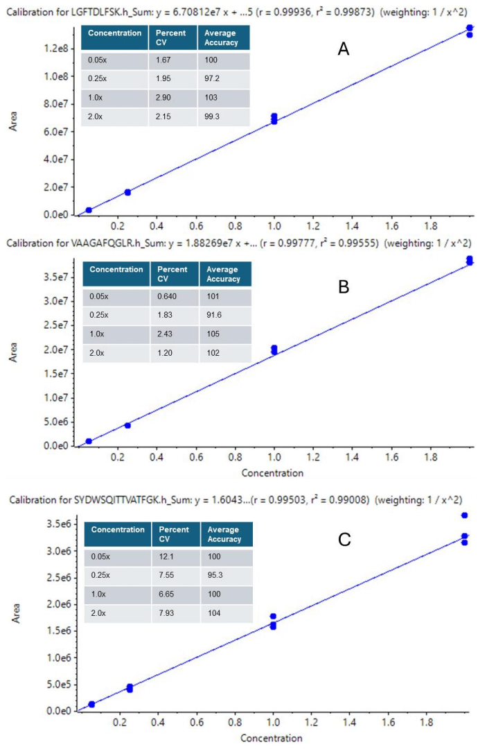
Response curves were measured across a concentration range spanning heavy-labeled peptide spike-in ratios of 0.05x to 2x the recommended PQ500 to plasma spike-in ratio. The linearity for the relative quantitation was assessed for each peptide individually from these curves. Figure 4 shows the curves for three peptides across 1.5 orders of peak area response. The low CVs and excellent accuracies around the spike-in ratio for the heavy peptide standards show that this method enables accurate quantitation of native unlabeled peptides across various concentration ranges and provides confidence in measuring plasma protein concentrations.
Plasma protein concentrations of biomarker candidates are shown in Table 3 as examples of the quantitative capabilities for the SCIEX 7500+ system. In the first example, the plasma protein concentration for Kallistatin, a serine proteinase inhibitor being studied as a potential marker for pneumonia4 and inflammation after kidney transplant5 was evaluated. While the concentration (mg/L) determined for Kallistatin was lower than what is reported for LC-MS-based analysis in the Human Protein Atlas8 and literature5, immunoaffinity-based determined values reported in the literature ranged from 5.3 to 82.7 mg/L for healthy individuals4. In the next example, Leucine-rich alpha-2-glycoprotein (LRG1), a potential Leukemia biomarker, was found at a concentration significantly lower compared to the value reported in the Human Protein Atlas. However, the measured value from the SCIEX 7500+ aligned with the literature reported mean value of 2.03 mg/L in normal plasma, measured by LC-MS6. In the last example, Di-N-acetylchitobiase, a protein involved in the degradation of N-linked glycoproteins that has been studied as a potential biomarker for oral cancer7, was found at a concentration 4x higher than the LC-MS-based plasma concentration reported in the Human Protein Atlas or literature9.

Conclusions
- The SCIEX 7500+ system enables MRM quantitation of large peptide panels using MRM dwell times as low as 1.5 ms with excellent peak area precision, accuracy, and signal-to-noise ratios
- >97% of 709 labeled human plasma peptides were quantified with CVs <10% and a median CV of 2.5%, using a 20-minute microflow LC gradient
- M icroflow LC methods (using a 0.15 mm ID column at a flow rate of 1.5 uL/min) provide sensitivity >5-fold higher than analytical flow LC
References
- Smith, Richard D (2012). Mass Spectrometry in Biomarker Applications: From Untargeted Discovery to Targeted Verification, and Implications for Platform Convergence and Clinical Application. Clinical Chemistry, Volume 58, Issue 3, Pages 528–530 https://doi.org/10.1373/clinchem.2011.180596
- Chen, Z., et.al. (2021). In-depth site-specific analysis of N-glycoproteome in human cerebrospinal fluid and glycosylation landscape changes in Alzheimer's disease. Mol & Cell. Proteomics, 20:100081.
- Mclean, Brendan et al. (2010). Skyline: an open source document editor for creating and analyzing targeted proteomics experiments. Bioinformatics Apr 1;26(7):966-8. https://doi.org/10.1093/bioinformatics/btq054
- Wei-Chieh Lin et al. (2013) Plasma kallistatin levels in patients with severe community-acquired pneumonia. Crit Care. 17(1):R27. 10.1186/cc12507
- Sofia Nteak et al. (2024). Longitudinal fluctuations in protein concentrations and higher-order structures in the plasma proteome of kidney failure patients subjected to a kidney transplant. J Proteome Res Jun 7;23(6). 10.1021/acs.jproteome.4c00064
- Chin Hin Ng et al. (2017) Plasma LRG1 Protein Are Overexpressed in Acute Myeloid Leukemia with High Sensitivity and High Specificity in Differentiating Leukemic Plasma from Normal Plasma — a Potential Biomarker for Minimal Residual Disease Detection. Blood 130 (Suppl_1) : 5083. http://doi.org/10.1182/blood.V130.Suppl_1.5083.5083
- Qiao Peng et al. (2023) The potential roles of cigarette smoke-induced extracellular vesicles in oral leukoplakia. European Journal of Medical Research 28:250. https://doi.org/10.1186/s40001-023-01217-0
- The Human Protein Atlas. https://www.proteinatlas.org/
- Claudia Escher et al. (2014) Determination of the variability and normal ranges of biomarker proteins in human plasma from healthy donors. https://biognosys.com/media/biognosysplasma-proteins-abs-quantcase-study.pdf