Abstract
This technical note describes a streamlined workflow for the in vitro characterization of liraglutide peptide catabolites using the ZenoTOF 8600 system. By leveraging complementary CID and EAD fragmentation, the workflow delivers enhanced sequence coverage and greater confidence in structural assignment, enabling more comprehensive characterization of peptide catabolites (Figure 1).
Synthetic therapeutic peptides represent a growing class of biotherapeutics. GLP-1 receptor agonists are modified versions of native human GLP-1, transforming the treatment landscape for type 2 diabetes mellitus (T2DM), obesity, and weight loss management.1 Characterization of peptide catabolites across diverse masses and abundance levels remains challenging due to limited MS/MS sensitivity from TOF duty cycle constraints. In addition, CID fragmentation is often dominated by b and y ions, resulting in incomplete sequence coverage, ambiguous localization of modifications or cleavage sites, and reduced confidence in catabolite assignment.2
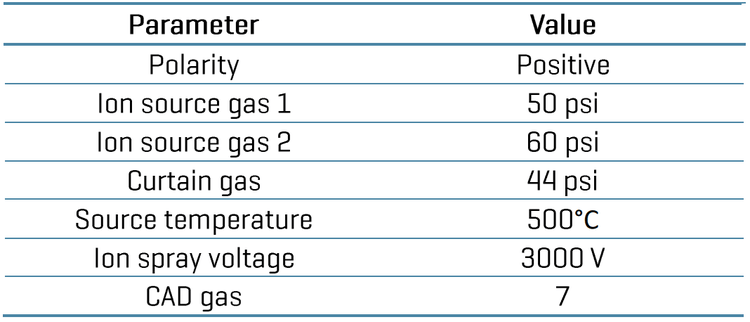
The ZenoTOF 8600 system enhances MS/MS sensitivity with Zeno trap3, while EAD provides complementary c′ and z•ion information beyond traditional CID fragmentation.4 This combination increases sequence coverage and confidence in assigning peptide catabolites, including low-level metabolites.
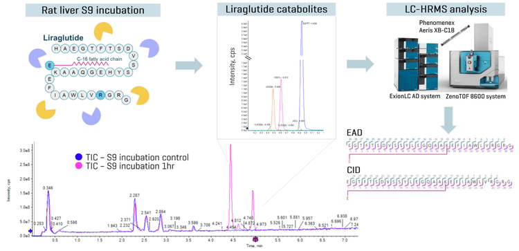
Key benefits for catabolite identification using ZenoTOF 8600 system
- Enhanced structural confidence: CID and EAD deliver complementary fragment ions that expand sequence coverage and strengthen confidence in liraglutide catabolite assignment.
- Improved duty cycle and sensitivity: The Zeno trap improves TOF duty cycle, increasing MS/MS sensitivity for confident identification of low-level peptide catabolites.
- Streamlined workflow: Improve insights into structure-metabolic stability relationships by leveraging an end-to-end workflow unifying data acquisition to processing on a single LC-HRMS platform.
Introduction
Comprehensive elucidation of drug metabolism pathways is critical for assessing drug safety and therapeutic efficacy as catabolites can contribute to pharmacological activity, toxicity, and drug–drug interactions. Accurate identification and structural characterization of peptide catabolites are therefore essential components of drug metabolism and pharmacokinetic (DMPK) studies.
Synthetic therapeutic peptides represent an important and growing class of biotherapeutics as their structures can be engineered to improve target specificity, metabolic stability , and pharmacokinetic properties. This technical note uses liraglutide, a GLP-1 receptor agonist, as a model compound. Liraglutide is a modified analog of native human GLP-1 designed to preserve GLP-1 receptor activity while overcoming the short half-life of the endogenous peptide, which is rapidly degraded by enzymes such as dipeptidyl peptidase-4 (DPP-4) and neutral endopeptidase (NEP). 5 Structurally, liraglutide contains a Lys34-to-Arg substitution and a C16 fatty acid attached to Lys26 through a glutamic acid spacer. These modifications improve enzymatic stability, promote plasma protein binding, and extend systemic exposure.
However, the same structural modifications that improve therapeutic performance also increase the complexity of catabolite identification. Peptide catabolism often involves enzymatic backbone cleavage, producing related catabolites that can differ by only a few residues, charge states, or structural modifications. For lipidated peptides such as liraglutide, confident assignment requires not only identifying peptide cleavage sites but also determining whether key structural features, such as the lipidated side chain, are retained or lost.
LC-HRMS has become a central tool for DMPK studies because accurate-mass detection, isotope pattern recognition, and high-resolution MS/MS fragmentation provide the structural information needed to assign peptide catabolites and localize cleavage sites. Commonly used CID-based fragmentation provides useful peptide structural information but can be limited by incomplete or uneven sequence coverage. CID spectra are often dominated by b- and y-type ions, and some regions of the peptide sequence may fragment poorly, making it difficult to confidently localize cleavage sites or distinguish closely related catabolites.2
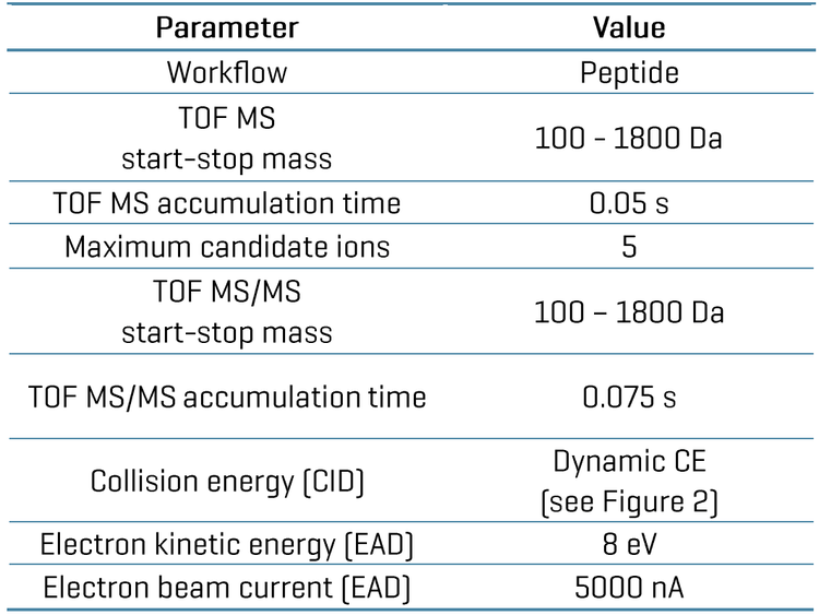
The ZenoTOF 8600 system addresses these challenges by combining high-resolution accurate-mass detection, enhanced MS/MS sensitivity through the Zeno trap, and complementary fragmentation using EAD.3,4 EAD provides an additional fragmentation approach that uses tunable electron kinetic energy to generate complementary backbone fragment ions, including c ′ and z•ions, expanding sequence coverage beyond CID alone.
By combining CID and EAD fragmentation, this workflow provides more comprehensive structural information for liraglutide catabolites, improving confidence in sequence assignment, cleavage-site localization, and characterization of lipidated catabolites in biological matrices. The observed liraglutide cleavage sites were consistent with reported GLP-1 catabolic pathways, supporting the biological relevance of the in vitro characterization workflow.5,6
Methods
Sample preparation for incubation in rat S9 fractions: Liraglutide at a 10 µM starting concentration was incubated in rat S9 fractions at 37°C for 1 hour. Samples were removed from the incubation and quenched with acetonitrile at a 2:1 (v/v) ratio. Samples were vortexed for 30 seconds and centrifuged at 12,000 rcf for 12 m ins at room temperature. Samples were diluted with 1:5 (v/v) with water containing 0.1% formic acid.
Sample preparation for enzyme incubation: Liraglutide at a 10 µM starting concentration was incubated in phosphate buffer (pH 7.4) containing 2 µg/mL DPP-4 or 2 µg/mL NEP at 37°C for 1 hour. Sample extraction and dilution were carried out as mentioned under “sample preparation for incubation in rat S9 fractions”.
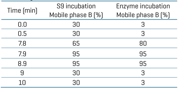
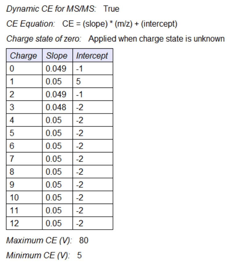
Chromatography: Sample separation was performed using an ExionLC AD system (SCIEX) at a 0.4 mL/min flow rate on a Phenomenex Aeris XB C18 (2.1 x 100 mm, 1.7 µm, 100 Å) column. A 10- minute gradient was run using 0.1% formic acid in water as mobile phase A and 0.1% formic acid in acetonitrile as mobile phase B. The conditions for mobile phase B are summarized for rat S9 incubation and enzyme incubation (Table 1). The column temperature was maintained at 40°C. An injection volume of 5 µL was used for analysis. A mixture of equal volumes of acetonitrile, methanol, and water was used as the needle wash solvent.
Identification of liraglutide peptide catabolites
Automated structural assignment was performed using Molecule Profiler software, where the ppm error for all fragment ions was observed to be <10 ppm, enabling confident identification of peptide catabolites.
Figure 3 shows a summary of the liraglutide catabolite investigated in rat S9 fraction incubations. Among 16 catabolites detected with charge states spanning +2 to +4, 2 examples with the highest peak area were highlighted in this technical note. For all the selected catabolites, t he fatty acid chain was observed to be intact. All selected catabolites were reported in the literature.5
To further confirm the catabolite assignments observed in rat S9 incubations, liraglutide was incubated with DPP-4 and NEP enzymes in phosphate buffer. Comparison of the enzymatically generated catabolite profiles with those obtained from rat S9 incubations provided orthogonal confirmation of the proposed biotransformation pathways as shown in Figure 4.
Figure 5 shows one of the catabolites of NEP driven digestion product M41-1 (m/z 790.1, charge state of +2). CID based sequence coverage was incomplete due to the poor fragmentation close to the C-terminal of the peptide, whereas EAD based z ions (such as m/z 216.1224, m/z, 587.3174) provided fragment information that resulted in improvement in the sequence coverage.
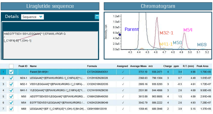
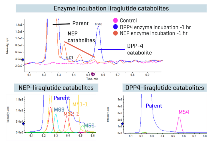
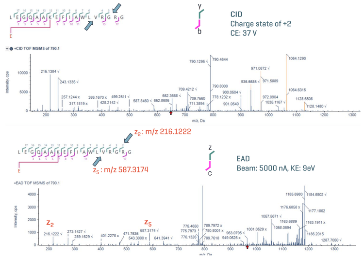
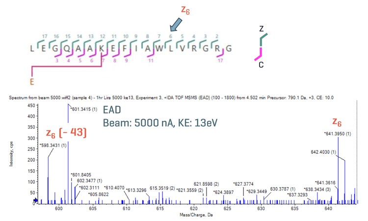
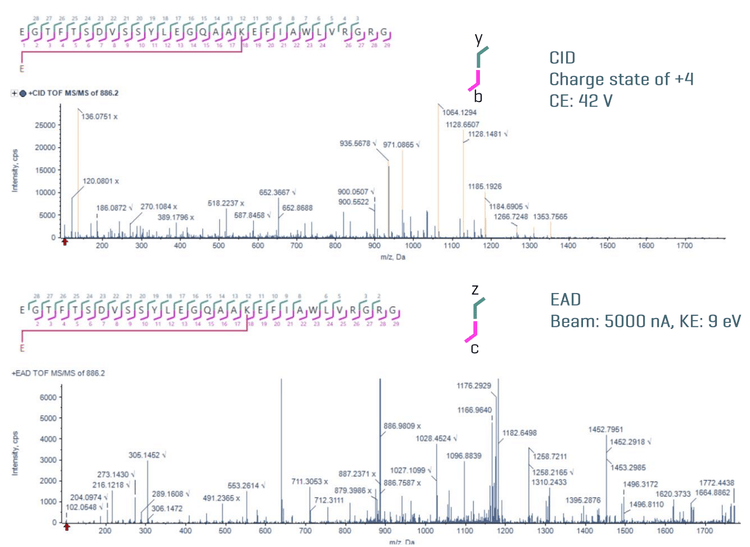
Figure 6 shows a z-43 fragment ion (m/z 598.3431) generated by EAD, indicative of Leu. Figure 7 shows the CID and EAD spectra for the dominant catabolite M54 (m/z 886.2, charge state of +4) produced by DPP- 4 digestion.3 Both CID and EAD fragments provide full sequence coverage of M54 through complementary fragmentation pathways. A near-complete set of b and y ions from CID and c′ and z•ions from EAD enabled extra confidence in structure characterization and assignment of the catabolite.
In summary, CID and EAD spectra both provided complete sequence coverage of peptide catabolites. In addition, EAD-based fragmentation provides additional c′ and z•ions, facilitating enhanced confidence in catabolite identification.
Conclusions
- More comprehensive sequence coverage was achieved by combining complementary CID and information-rich EAD fragmentation on the ZenoTOF 8600 system.
- The identification of catabolites in a wide concentration range with varying charge states was easily achieved with the Zeno trap, enabling enhanced MS/MS sensitivity for both EAD and CID applications.
- Fragments from EAD and CID spectra were identified and processed in a single result file using Molecule Profiler software to achieve more accurate structure assignment of catabolites.
- Informative data was generated using a quick and easy-to-use workflow on a streamlined platform, accelerating the early drug discovery process.
References
- Nauck MA, Quast DR, Wefers J, Meier JJ. GLP-1 receptor agonists in the treatment of type 2 diabetes - state-of-the-art. Mol Metab. 2021 Apr;46:101102.
- Martin DB, Eng JK, Nesvizhskii AI, Gemmill A, Aebersold R. Investigation of neutral loss during collision-induced dissociation of peptide ions. Anal Chem. 2005 Aug 1;77(15):4870-82
- Tunable Electron-Assisted Dissociation (EAD) MS/MS to preserve particularly labile PTMs. SCIEX technical note, RUO-MKT-02-13006-B.
- Enhanced drug catabolite identification using collision-induced dissociation (CID) and electron-activated dissociation (EAD). SCIEX technical note, MKT-29204-A.
- Monika Malm- Erjefält, Inga Bjørnsdottir, Jan Vanggaard, Hans Helleberg, Uffe Larsen, Berend Oosterhuis, Jan Jaap van Lier, Milan Zdravkovic, Anette K. Olsen,Metabolism and Excretion of the Once-Daily Human Glucagon-Like Peptide-1 Analog Liraglutide in Healthy Male Subjects and Its In Vitro Degradation by Dipeptidyl Peptidase IV and Neutral Endopeptidase, Drug Metabolism and Disposition, Volume 38, Issue 11, 2010
- Sharma R, McDonald TS, Eng H, Limberakis C, Stevens BD, Patel S, Kalgutkar AS. In vitro metabolism of the glucagon-like peptide-1 (GLP-1)-derived catabolites GLP-1(9-36)amide and GLP-1(28-36)amide in mouse and human hepatocytes. Drug Metab Dispos. 2013 Dec;41(12):2148-57
- Differentiation of leucine and isoleucine for enhanced sequence variant analysis using electron activated dissociation. SCIEX Technical Note, MKT-30799-A.
- Characterization of adeno associated virus capsid proteins using orthogonal intact MS, top-down MS/MS, and peptide mapping workflows. SCIEX Technical Note, MKT-38462-A.