Analysis of post-translational modifications using fast electron-activated dissociation (EAD)
Using the ZenoTOF 7600 system with Zeno MS/MS
Nick Morrice1, Alexandra Antonoplis2 and Christie Hunter2
1SCIEX, UK; 2SCIEX, USA
Abstract
Electron activated dissociation (EAD) is a fragmentation mode that involves capture of electrons by molecular ions to form radicals that dissociate into fragment ions. EAD often preserves labile post translational modifications (PTMs) and provides peptide sequence information that is complementary to collision induced dissociation (CID). This work focuses on localization of peptide phosphorylation and glycation sites using EAD in a bottom-up liquid chromatography-mass spectrometry workflow.
Introduction
Characterization of human cellular proteomes enables the identification of disease biomarkers and new therapeutic targets. Mass spectrometry-based techniques, such as bottom-up proteomics, are widely used to analyze cellular proteomes. During bottom-up proteomics experiments, intact proteins are cleaved by enzymes to yield peptides and the resulting peptide sequences are determined by liquid chromatography-mass spectrometry (LC-MS/MS).
Several factors influence peptide MS/MS spectral data quality and depth of sequence coverage, including the data acquisition speed and fragmentation mode implemented. Data-dependent acquisition (DDA) is often used to analyze protein digests for protein identification. MS/MS sensitivity is a crucial performance attribute in DDA workflows. The ZenoTOF 7600 system uses the Zeno trap to increase the duty cycle to ≥90% across the entire fragment ion mass range, resulting in average signal gains of 5- to 6-fold in MS/MS mode for peptides.1-2 These gains enable significant improvements in proteins identified using DDA workflows.3
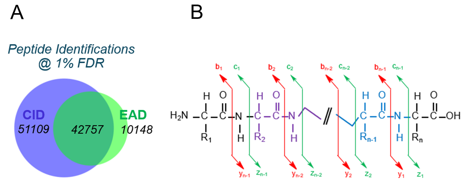
For proteomics experiments, 2 fragmentation options are available on the ZenoTOF 7600 system: collision-induced dissociation (CID) and electron-activated dissociation (EAD) (Figure 1). Unlike CID, which relies on the collision of ions with nitrogen gas to fragment molecules at their weakest bonds, EAD involves the capture of electrons by molecular ions to form a radical state that fragments. CID and EAD result in different fragments for the same peptide, with CID yielding b and y ions and EAD yielding additional c’ and z• ions. EAD fragmentation provides complementary sequence information to CID and often preserves post-translational modifications (PTMs) that undergo neutral loss in a CID experiment.4-5 Many different modifications can be labile during MS analysis, including phosphorylation, sulfation and O-GlcNAc modifications. This work focuses on DDA method development and analysis of HeLa digest using EAD, with an emphasis on site-specific localization of phosphorylation sites. In addition to phosphorylated peptides, glycated human peptides were also investigated using EAD.
Figure 1. Comparison of peptide identifications at 1% FDR as a function of fragmentation mode on the ZenoTOF 7600 system. A) Due to the higher frequency of sampling with CID, peptide identifications were approximately 2-fold higher than EAD with this fragmentation mode but EAD provided over 10,000 new peptide identifications. B) Illustration of typical fragment ions produced by CID (marked with red lines) and EAD (marked with green lines).
Key features of the ZenoTOF 7600 system for Zeno EAD DDA analysis
- The ZenoTOF 7600 system supports collision-induced dissociation (CID) and electron-activated dissociation (EAD), which can provide complementary peptide sequence information for routine proteomic analysis
- The use of the Zeno trap yields a 5- to 6-fold gain in MS/MS sensitivity for peptides, enabling the identification of more high-confidence proteins and peptides.2-3
- Zeno EAD DDA methods using microflow chromatography were developed for sensitive analysis of HeLa digest
- EAD complements CID by providing a fast, orthogonal fragmentation strategy that can identify additional peptides
- EAD is easy to implement for DDA workflows in SCIEX OS software with automated EAD tuning and streamlined method building
Methods
Sample preparation: A 100 µg sample of digested HeLa cell lysate was fractionated into 44 samples using high pH RP-HPLC, as previously described.6 Then, 10% of each fraction was injected for LC-MS/MS analysis. For analysis of glycated peptides, human serum albumin was used from human plasma.
Chromatography: Peptide digests were separated using a Waters ACQUITY UPLC M-class system with a flow rate of 6 µL/min in direct-inject mode. A Phenomenex Kinetex 2.6 µm XB-C18 LC column (100 Å, 150 x 0.3 mm) was used. A rapid linear gradient from 5–30% B over 21 minutes was used to interrogate the fractions. Mobile phase A was water with 0.1% formic acid and mobile phase B was acetonitrile with 0.1% formic acid.
Mass spectrometry: A ZenoTOF 7600 system that was equipped with the OptiFlow Turbo V source7 using a low microflow probe and electrode was used for all data acquisition. DDA parameters that were implemented for all experiments included a TOF MS accumulation time of 250 ms and an exclusion time of 6 seconds. Only precursors with charge states in the range of 2-5 with intensities greater than 100 cps were selected for fragmentation. For CID acquisition, the maximum number of candidate ions per cycle was 45 and the accumulation time was 20 ms. For EAD analysis of HeLa digest, the maximum number of candidate ions was 10 per cycle and an accumulation time of 50 ms was used. The reaction time for EAD was 20 ms with an electron beam current of 3000 nA with a KE of 0 eV. For analysis of glycated peptides, 20 candidate ions were used per cycle with a 20 ms reaction time and 25 ms total acquisition time.
Data processing: Mascot software (Matrix Science, version 2.6) was used for all data processing. Data files were searched against the Swissprot_Human database using the SCIEX EAD algorithm for EAD and the ESI-QUAD-TOF algorithm for CID. For analysis of phosphorylated peptides in HeLa digest, the following modifications were searched in Mascot software: fixed: carbamidomethyl; variable: phosphorylation (on serine, threonine and tyrosine); methionine oxidation and n-terminal acetylation. Scaffold software (Proteome Software Inc., version 4.8.5) was used to confirm MS/MS-based peptide and protein identifications. For analysis of glycation sites, hexose variable modifications were searched in Mascot software with a fixed carboxymethyl modification. For both phosphorylated and glycated peptides, figures were generated using Bio Tool Kit in SCIEX OS software.7
Determining Zeno EAD DDA parameters for proteomic analysis of labile PTMs
In order to select the acquisition settings for Zeno EAD DDA analysis of HeLa digest, a variety of parameters were tested, including electron beam current, electron kinetic energy, reaction time and accumulation time. For peptides, it was determined that using an electron KE of 0 eV and a beam current of 3000 nA promoted fragmentation and yielded high fragment ion intensities.
A reaction time of 20 ms coupled with an accumulation time of 50 ms enabled 2 EAD reactions to be performed for each MS/MS spectrum and these parameters were employed for all experiments. The Zeno trap was turned on to improve fragment ion sensitivity, as Zeno trap activation results in a restoration of duty cycle to ≥90% across the entire mass range of the MS/MS spectrum.1
For analysis of glycated peptides, further optimization was performed and it was determined that using a method with 20 MS/MS candidates and a 20 ms EAD reaction time yielded accurate identification of challenging modifications.
Proteomic coverage of HeLa digest obtained using EAD
Forty-four fractions of HeLa digest were separated using a fast 21-minute gradient and analyzed with DDA. Both CID and EAD MS/MS fragmentation were evaluated and results were processed and visualized using Mascot software and Scaffold software. In the CID experiment, 93,866 peptides were identified at 1% FDR using Mascot software as the database search engine (Figure 1). This search was performed using the standard settings for CID fragmentation and included 1 fixed and 5 variable modifications in the search space.
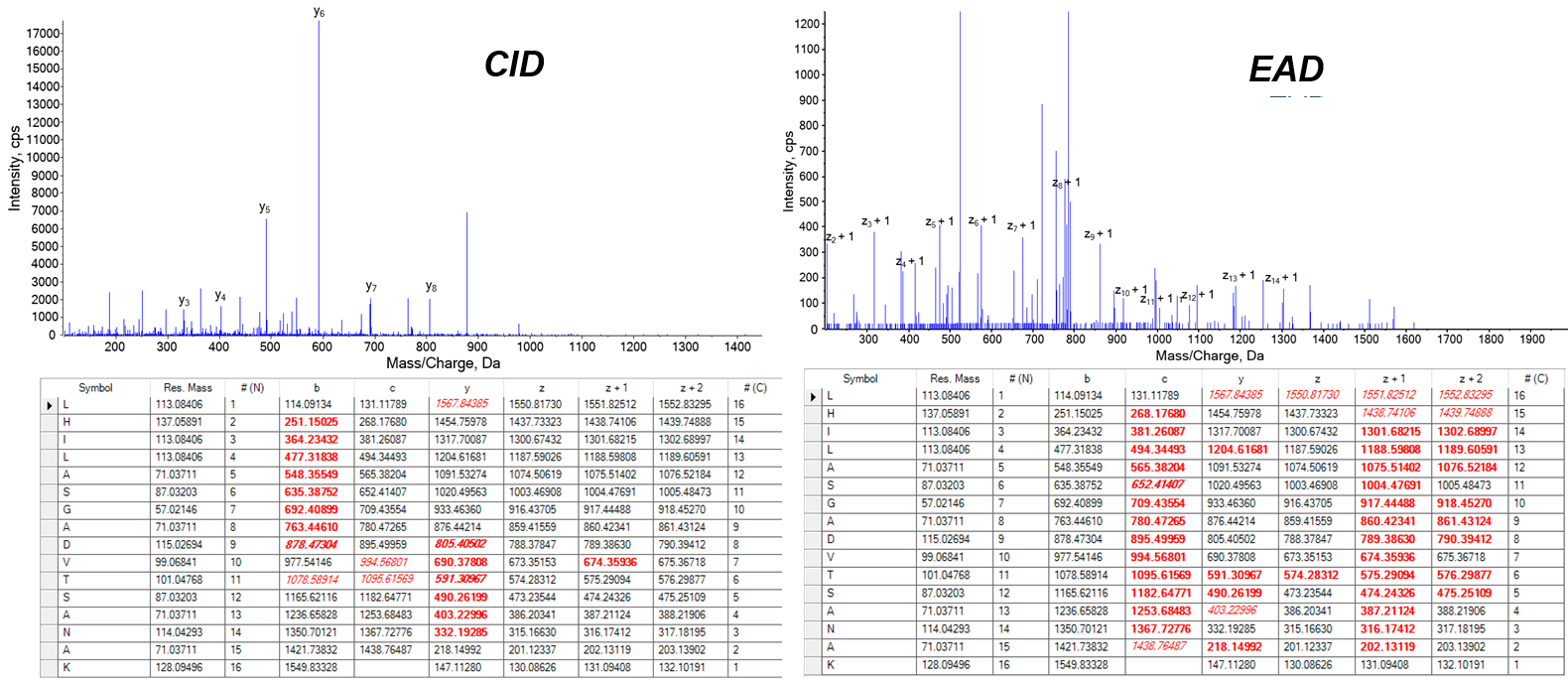
When the fragmentation mode was switched to EAD, 52,905 peptides were identified at 1% FDR (Figure 1). Fewer total numbers of peptides were identified using EAD due to the slightly slower acquisition speed (2.5x slower). However, the orthogonal fragmentation mode provided complementary sequence information (Figure 2) and identifications of peptides not found using CID, yielding 11% more peptides. This highlights that EAD is able to run at a frequency compatible with the LC timescale and with DDA.
Figure 2. Comparing CID and EAD fragmentation of a non-phosphorylated peptide. (Top) CID fragmentation of the peptide, DSYVGDEAQSKR, yields y and b ions for peptide sequencing, with detected fragment ion masses (+1 charge state) shown in bold red in the table beneath the spectrum. Red italics indicate a charge state other than +1. (Bottom) Nearly complete z• (z+1 in table) and c’ ion series were detected for the same peptide when fragmented by EAD. Together, CID and EAD provide complementary information for sequencing peptides.
Phosphorylation site localization using Zeno EAD MS/MS spectra
Although the HeLa cell digest was not specifically prepared to preserve PTMs, 157 phosphopeptides were identified at 1% FDR with EAD fragmentation. The raw phosphopeptide MS/MS spectra were evaluated in Explorer in SCIEX OS software. Using the Bio Tool Kit, the peptide sequences could be overlaid on the spectra to evaluate the specific fragments and the fragmentation coverage (Figures 3-5).7
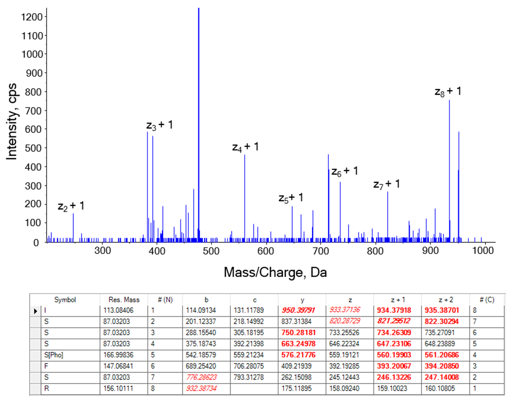
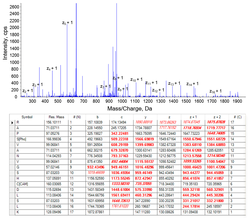
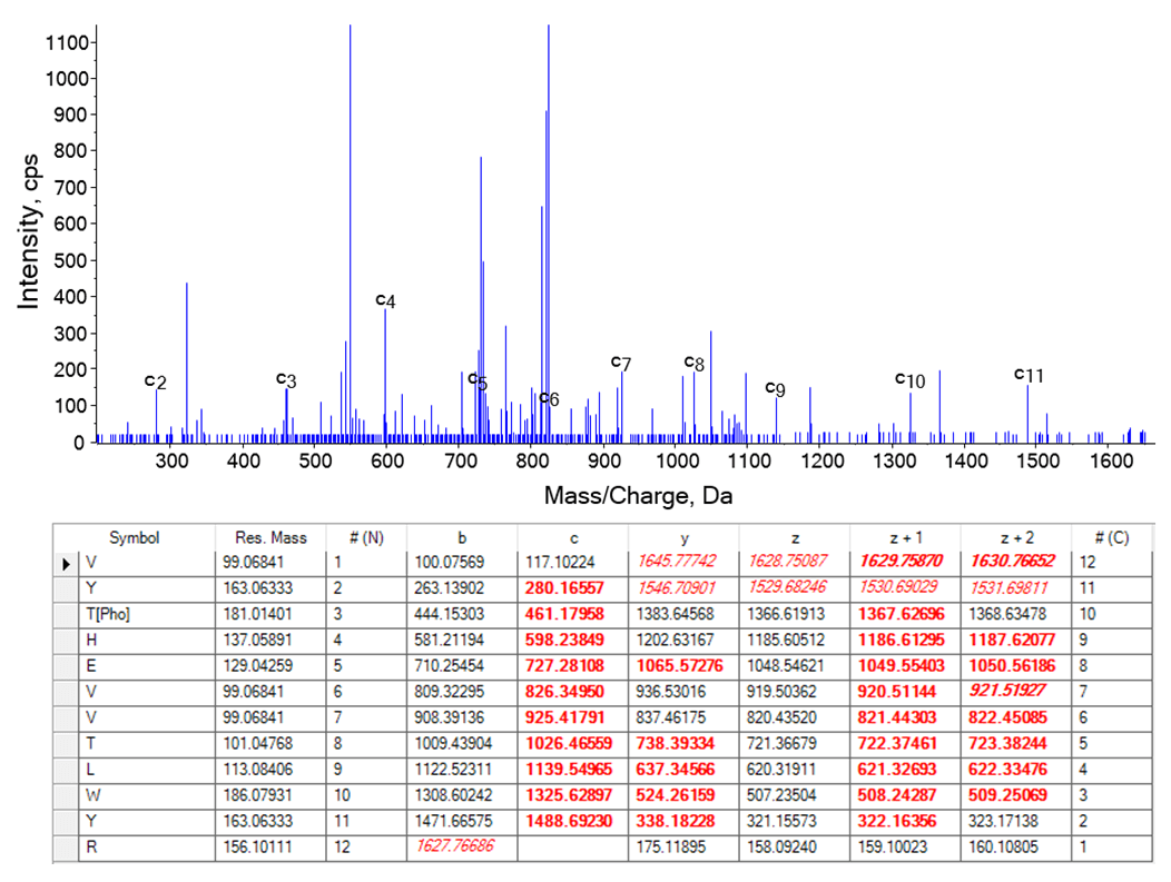
EAD spectra can be used to identify the sequence and phosphorylation site in peptides, including peptides with multiple serines (Figure 3). Other phosphorylation sites were located using EAD spectra, including a site on a 1889.88 Da peptide with 17 amino acids (Figure 4) and a site on a threonine residue near the peptide N-terminus (Figure 5). The associated c’ and z• (z+1) ion series clearly detailed the location of the phosphorylation sites on the peptides.
Figure 3. Localization of phosphorylation site in a peptide with multiple serines. An EAD spectrum for peptide sequence ISSSS[Pho]FSR contains a nearly complete z• (z+1) ion series. The associated fragment ion masses are shown in the table below the spectrum for reference.The modification site was localized with 97.42% probability in Mascot software. Bold red indicates the fragments found with +1 charge state and red italics indicate fragments found with a different charge state.
Figure 4. Identification of phosphorylation site in a 17-residue peptide. The presence of c’ and z• (z+1) fragment ions enable the identification of a phosphorylated serine in the sequence of a 1889.88 Da peptide. The modification site was located with 100% probability in Mascot software. Detected fragment ions (+1 charge state) are tabulated in bold red, with red italics indicating a charge state other than +1.
Figure 5. Localization of a phosphorylation site near the N-terminus of a peptide with tyrosine and threonine. The c’ fragment ion series enables localization of a phosphorylated threonine adjacent to a tyrosine. The c’ ion series is shown in the spectrum and the modification site was located with 99.98% probability in Mascot software. Both c’ and z• (z+1) ion series are highlighted in the table below the EAD spectrum and the detected fragments (+1 charge state) are highlighted in bold red.
Glycation site localization using Zeno EAD MS/MS
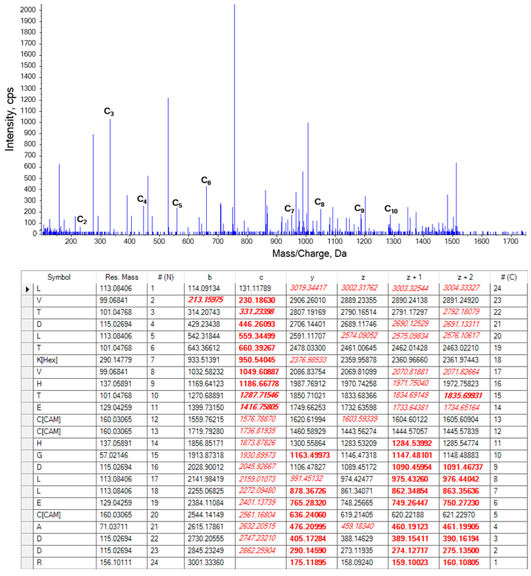
A panel of human peptides was analyzed to evaluate hexose and modifications using Zeno EAD DDA. While a hexose modification could not be localized using CID, EAD enabled unambiguous localization of a hexose modification on a lysine residue on a long, 24 amino acid peptide (Figure 6).
Figure 6. EAD fragmentation of a glycated peptide from human serum albumin protein. A hexose modification was localized on a lysine using a nearly complete c’ ion series. Detected fragment ion masses are shown in red in the table below the EAD spectrum. The hexose modification was located with 100% probability in Mascot software. Bold red indicates the fragments found with +1 charge state, while the red italics indicate fragments found with a different charge state.
Conclusions
The Zeno EAD DDA workflow developed in this work enabled the robust identification of thousands of unique peptides in HeLa digest. High pH fractionation of HeLa digest followed by LC-MS/MS analysis with the ZenoTOF 7600 system generated a large-scale Zeno EAD DDA dataset for interrogation. Microflow chromatography was used to improve sensitivity and the Zeno trap was activated to both improve MS/MS sensitivity and yield high fragment ion intensities. This work highlights that Zeno EAD is fast and compatible with DDA experiments.
EAD provides an alternative fragmentation option for the study of proteomic samples and enables better characterization of some types of peptides, such as long tryptic peptides and peptides containing labile PTMs, compared to CID. In the EAD spectra evaluated here, intact z• (z+1) and c’ ion series were readily detectable for sequencing and localization of phosphorylation and glycation sites.
References
- Qualitative flexibility combined with quantitative power. SCIEX technical note, RUO-MKT-02-13053-A.
- Large-scale, targeted, peptide quantification of 804 peptides with high reproducibility, using Zeno MS/MS. SCIEX community post, RUO-MKT-18-13346-A.
- Over 40% More Proteins Identified with Zeno MS/MS. SCIEX technical note, RUO-MKT-02-13286-B.
- Tunable Electron-Assisted Dissociation (EAD) MS/MS to preserve particularly labile PTMs. SCIEX technical note, RUO-MKT-02-13006-B.
- Peptide fragmentation using electron activated dissociation (EAD). SCIEX community post, RUO-MKT-18-13296-A
- Large scale protein identification using microflow chromatography on the ZenoTOF 7600 system. SCIEX technical note, RUO-MKT-18- 14415 -A.
- Single source solution for low flow chromatography. SCIEX technical note, RUO-MKT-02-9701-A.
- Using Bio Tool Kit for interpreting peptide MS/MS data obtained using electron activated dissociation (EAD). SCIEX community post, RUO-MKT-18-13296-A.
 Click to enlarge
 Click to enlarge
 Click to enlarge
 Click to enlarge
 Click to enlarge
 Click to enlarge